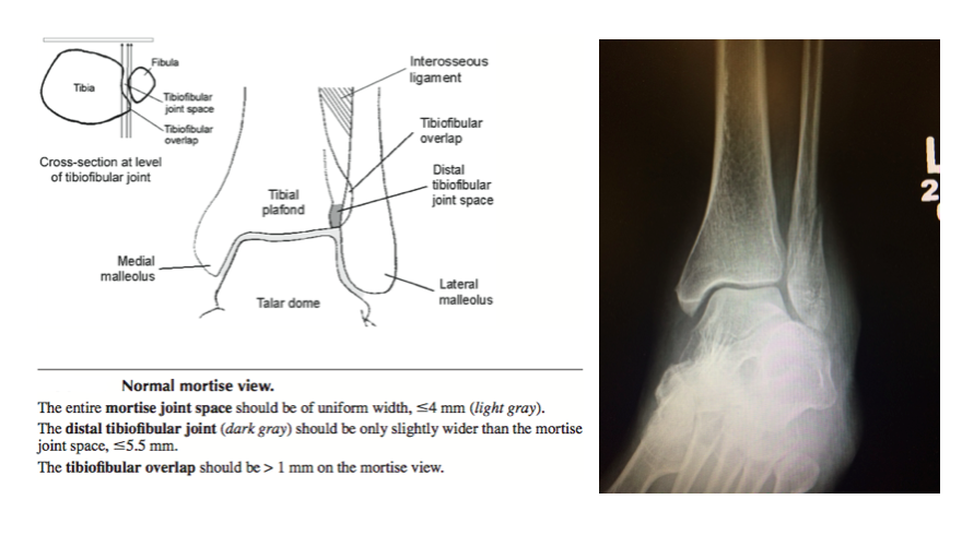
How do you code for Stress X Rays for the ankle. An ankle stress view should be interpreted using the same parameters as the mortise view return to that section of the post if you need a refresher.

Stress View Of Ankle With Deltoid Ligament Tear Radiology Case Radiopaedia Org
Of course the body part that needs stressing is usually hurting so oftentimes we make the doctor that ordered it do the holding.
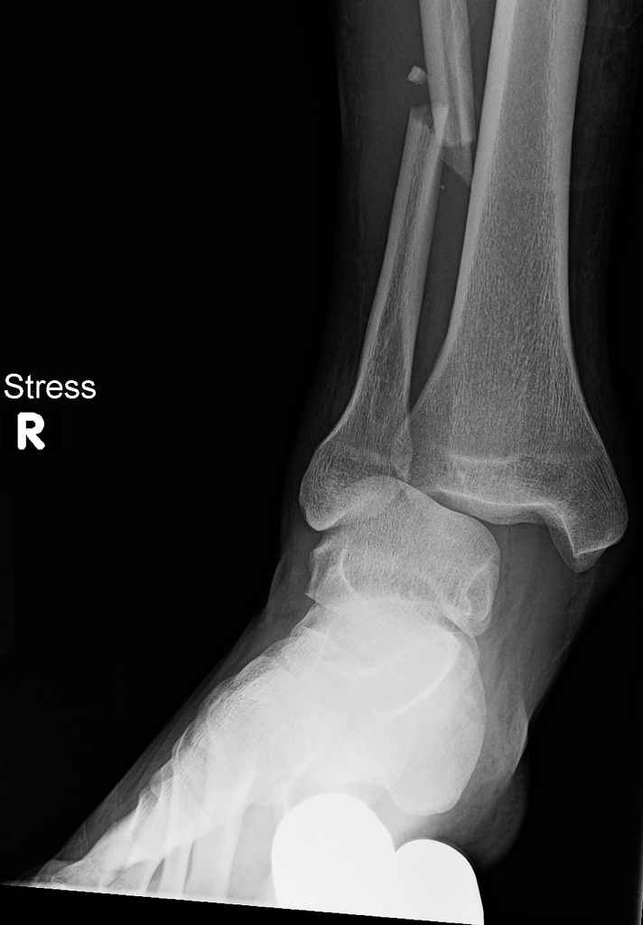
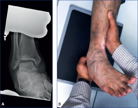
Stress x ray ankle. When performing this projection patients watchers or the physician must be present to hold the foot and ankle or the patient may hold and pull the strap looped around the foot. Medial clear space measurements were recorded. For X ray of the ankle.
Stress radiography is the gold standard for analyzing integrity of the capsuleligamentous structures. Theyre generally used to view the instability of a ligament or maybe an avulsion fracture in an ankle not too uncommon unfortunately. Consider additional views such as a full-length tibiafibula radiograph and imaging of the joint above and below ie.
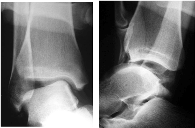
Patient position mechanical stress view the patient may be supine or sitting upright with the leg straightened on the table. In intermediate ankle injuries that have no syndesmotic widening on x-ray yet a high suspicion of injury will warrant a stress view to demonstrate dynamic widening of the ankle joint 1. You can see that the medial clear space has widened and it is in fact 4mm.
The ankle x-ray is used primarily to demonstrateexclude a fracture. In some cases a weight-bearing or a stress radiograph gravity stress or manual stress may also be required. A standard series includes an anteroposterior AP image a Mortise image and a lateral image.
Clinicians often use the talar tilt TT and anterior drawer AD stress x-rays to diagnose acute or chronic mechanical ankle instability. Usually you need to avoid putting weight on your leg for approximately 6 weeks. The initial x-ray was reported as normal but a T2-weigthed gradient echo of the knee shows bone marrow edema in the proximal tibia indicating the presence of a stress fracture.
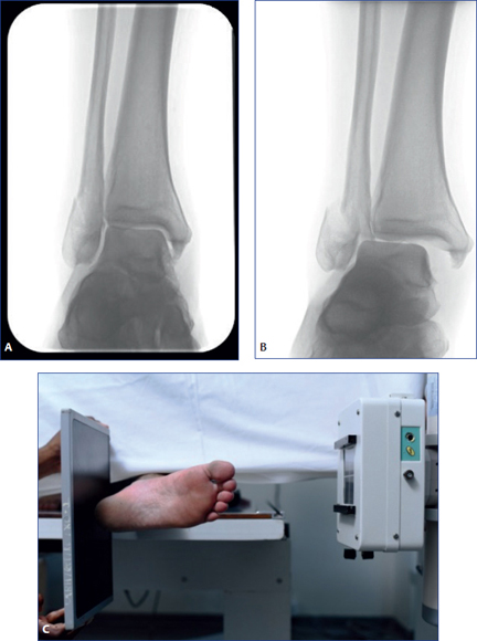
For the gravity stress view the patients were in a lateral decubitus position with their index ankle in neutral dorsiflexion and their ankle placed on a specially-designed table used for holding the leg and allowing the ankle to take a gravity-stress position. Stress radiography is used for the diagnosis and evaluation of trauma as well as disorders of ankle and midfoot. Its very simple actually.
When calcaneal pathology is suspected. Intra-operative stress radiography failed to detect approximately half. However the wide range of TT and AD values in normal and injured ankles makes interpretation of the test results difficult.
An ankle stress view should be interpreted using the same parameters as the mortise view return to that section of the post if you need a refresher. The fracture may be treated with a short leg cast or a removable brace. Manual external rotation stress and gravity stress tests were performed on injured and uninjured ankles of ankle fracture patients in a clinic setting.
AP Stress Ankle Joint X-ray Xray examination of ankle in AP stress projection includes Inversion and Eversion Position when performing this procedure proceed with utmost care with injured patient. Plain x-rays of the ankle will detect approximately half on AP view to two-thirds on mortise view of syndesmosis injuries. You will need to see your physician regularly for repeat x-rays to make sure the fracture does not change in position.
We regularly take stress views of various jointsusually the knee. Manual application stress performed by physician and 73620. A knee and a foot radiograph to rule out additional fractures.
Syndesmosis injuries frequently occur in association with tibial or fibular fractures. You can see that the medial clear space has widened and it is in fact 4mm. Conventional weight-bearing radiography of the foot and ankle stress radiographs can detect bone injuries concomitant ankle instability and foot deformities including tarsal coalitions.
In retrospect the sclerotic line on the x-ray also indicates the stress-fracture. A stress x-ray may be done to see if the fracture and ankle are stable. In painful conditions stress radiography can be done after injecting local anesthetic in the area of pain.
The X-ray cassette was held in a slot which rotated 15 from the vertical axis. Depending on the request various images can be made. Inversion Eversion.

Emrad Radiologic Approach To The Traumatic Ankle

The Varus And Valgus Stress Views And Anterior Drawer View In X Ray 4 Download Scientific Diagram

Ankle Stress Radiograph Of An 18 Year Old Female Patient Showing Varus Download Scientific Diagram

Ankle Fracture Stress View Radiographs Everything You Need To Know Dr Nabil Ebraheim Youtube

The Mortise Ankle X Rays After Deep Deltoid Ligament Transection Download Scientific Diagram
Radiology In Foot And Ankle Musculoskeletal Key

Lateral And Antero Posterior Ap Stress Ankle Radiographs Download Scientific Diagram

Emrad Radiologic Approach To The Traumatic Ankle
Ankle Stress Views Why When What Core Em

Ankle Stress Views Why When What Core Em

Gravity Stress View With Widening Of Medial Clear Space Download Scientific Diagram
Radiology In Foot And Ankle Musculoskeletal Key

Standard Stress Talar Tilt Angle Of The Right Ankle At The Preoperative Download Scientific Diagram
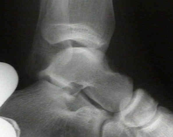
Radiographic Studies For Ankle Sprains Wheeless Textbook Of Orthopaedics
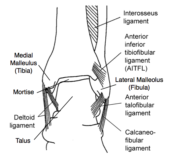
Ankle Stress Views Why When What Core Em
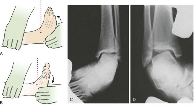
Imaging Of The Foot And Ankle Musculoskeletal Key
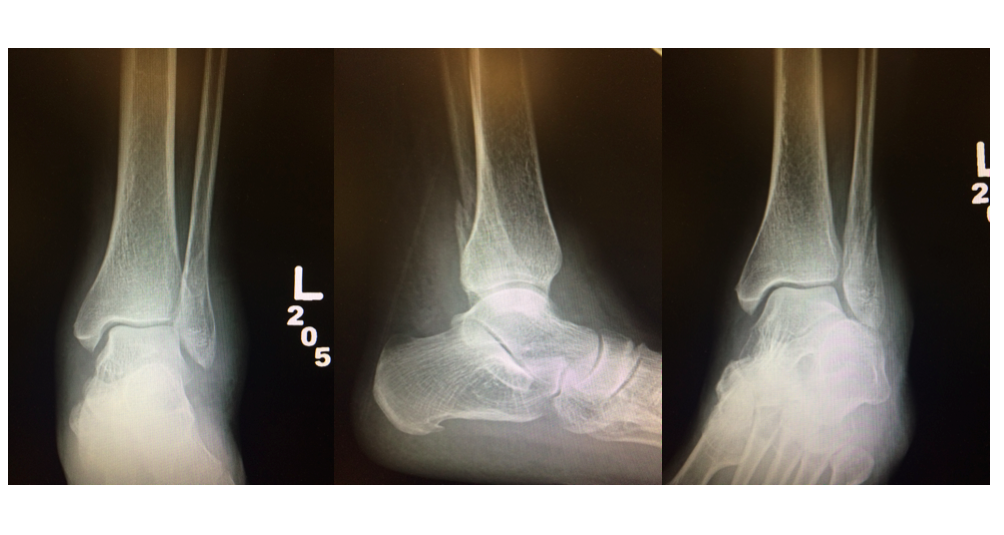
Ankle Stress Views Why When What Core Em

Unstable Ankle Injury Radiology Case Radiopaedia Org
0 comments